
“Selected Articles in English”
Selected articles originally published in Korean are now
translated into English,
making them accessible to the readers worldwide.
Prologue: The Journey Inward, Deciphering the Final Frontier
For thousands of years, humans have looked at the stars and wondered about the origin of the universe. Yet the most complex object in that universe sits right between our ears. The history of brain science is not just a list of dates and discoveries; it is the story of humanity finally turning its curiosity inward to ask: How do I think? How do I feel? And who am I?
The journey began with ancient civilizations debating whether our souls resided in our hearts or our heads. It progressed through the 19th century “spark” of electricity in a laboratory and the meticulous sketches of individual neurons that look like trees in a vast forest.
Today, we have entered a golden age. We no longer have to wait for an autopsy to see the brain’s secrets; we can watch it light up in real time as a person speaks, dreams, or solves a puzzle. As we move from simple observation to mapping the trillions of connections that make up the “Connectome,” we are realizing that brain science is the ultimate convergence of all human knowledge—from biology and physics to philosophy and artificial intelligence.
Welcome to the exploration of the “inner cosmos.” Whether you are a student, a curious reader, or a scientist, this journey belongs to everyone. After all, it is the brain’s greatest feat: the ability to study itself.
Neuroscience, or brain science, is the field of study dedicated to the brain and the entire nervous system. In the past, the term “neuroscience” was common during an era when neurons—the basic building blocks of the brain—were believed to be solely responsible for all brain activity. However, as it has been revealed that non-neuronal cells (glia) also play vital roles, the term “brain science” is now more universally used. In fact, there are actually more non-neuronal cells in the brain than neurons. Therefore, I will use the term “brain science” throughout the rest of this summary.
Introduction
The brain can be called the essence of human activity, as it is responsible for both our intelligence and our emotions. Naturally, humans—who are exceptionally curious compared to other animals—felt a drive to observe, analyze, and experiment on the brain. What’s fascinating, however, is that curiosity and research are themselves products of brain activity; in the end, it is the brain studying the brain. Regardless of this paradox, humans have been curious about the brain since ancient times, and brain science has steadily progressed through a longstanding desire to solve its mysteries. Let’s take a look at the history of this research to see how the scope of brain science began, expanded, and evolved.
Like any other science, the advancement of brain science involves a process of observation, analysis, and interpretation. And these activities are impossible without the development of research methodologies. In the early days, the invention of observation techniques and their integration into the field drove the initial progress of brain science. Later, the emergence of innovative technologies for analysis and interpretation led to groundbreaking advancements. Therefore, while explaining the history, I will also mention technologies that provided breakthroughs for brain research, even if they were not directly related at first.
Brain Science in Ancient Times
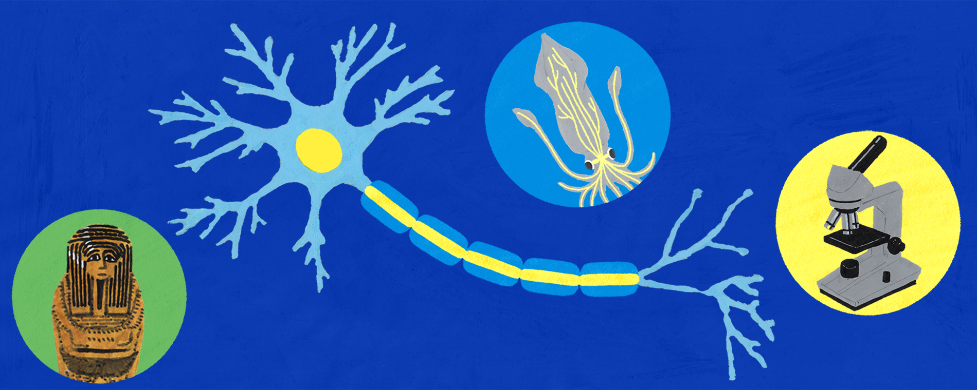
In ancient times, people believed the body was a vessel where the soul, mind, or spirit resided. Understanding which part of the body ancient humans believed housed the soul helps us gauge their level of understanding of the brain. As many of you know, when the ancient Egyptians made mummies, they broke the brain into pieces, extracted it through the nostrils, and threw it away. Organs believed to be necessary in the afterlife—such as the stomach, intestines, lungs, and liver—were stored separately in “Canopic jars.” There was no jar for the heart; it was left inside the mummy’s body. The Egyptians believed the spirit resided in the heart, so they thought a person could not be fully restored upon reincarnation without it. In reality, mummies don’t reincarnate, but even if they did, they would not be able to see, hear, or remember anything—because they would have no brain.
In ancient Greece, Hippocrates (c. 460–370 BC), often called the father of medicine, stated that “the brain is the place that governs human intelligence and emotions.” However, Aristotle (384–322 BC) claimed that “the brain is merely a cooling mechanism for blood or fluids heated by the excited heart, and the heart is where the mind resides.” This debate was eventually settled by the research of Claudius Galen (c. 129–199 AD), a Greek physician who served four Roman emperors. Galen performed human dissections and conducted extensive experimental research, particularly on the nervous system. Based on his dedicated work, he asserted that the brain controls both sensation and movement.
The Chinese Character for ‘Brain (腦)’
While precise records from ancient China are scarce, the existence of the Chinese character for “Brain” (腦) suggests that someone likely observed the organ. Looking at the character 腦, the left side is the radical for “flesh” (肉), and the right side features a symbol for an opening/crevice (凶) topped by “curvy lines” (巛) representing energy or hair. To use a bit of unscientific imagination, perhaps someone saw a corpse on a battlefield with a damaged skull, exposing the brain and told others that inside the skull was a “ghastly-looking, wrinkled lump of meat.” In reality, the character (腦) is an ideogram representing the “vital energy” (氣) rising to the head and the fontanelle (soft spot/opening) of the skull.
In summary, brain science in ancient Egypt and Greece was largely a philosophy based on the reflections and experiences of thinkers and physicians. It was not until the Roman era, when Galen began observing brain structures through dissection, that the field of morphology known as neuroanatomy was born. It is at this point that brain science truly began to emerge as a formal science. Since researchers of this time could only use their naked eyes, one can imagine how desperately they needed observation tools like microscopes.
Brain Science in the Middle Ages
The Middle Ages was a period when metaphysical reflections on the soul took precedence over scientific brain research due to religious constraints. Because of this, brain science during this era appeared to go through a “Dark Age,” with almost no official research records remaining. However, the story changes when we focus on the “witches” who frequently appear in folk tales and the “potions” they brewed. The secret formulas of these witches, known at the time for causing hallucinations or seizures, were likely based on sophisticated neuropharmacological knowledge.
The ingredients used in these potions can be categorized into plant-based and animal-based substances, both of which contain physiologically significant components from the perspective of modern medicine. In particular, plants from the nightshade (Solanaceae) family, such as Belladonna and Mandrake, contain powerful tropane alkaloids. These compounds act on receptors in the central nervous system to induce hallucinations, paralysis, and pain relief, or sometimes exhibit fatal toxicity.
A prominent example where such “nervous system contamination” led to a historical tragedy is the Salem Witch Trials of 1692 in Massachusetts, USA. Modern scientists propose ergot fungus (Claviceps purpurea) poisoning as one of the potential causes behind this infamous event. This fungus, which parasitizes rye and wheat, produces “ergot alkaloids,” which include lysergic acid derivatives—the chemical precursor to the powerful hallucinogen LSD. Ergot poisoning (ergotism) manifests in two distinct forms. The first is convulsive ergotism, which causes muscle spasms, seizures, a tingling sensation like insects crawling on the skin, extreme mental confusion, and hallucinations filled with terror. These symptoms align remarkably well with the abnormal seizures and testimonies of seeing the devil reported by the young girls in Salem at the time. The second is gangrenous ergotism, a condition where strong vasoconstriction cuts off blood supply to the limbs, causing the tissue to rot away. Medieval people called this “St. Anthony’s Fire” because patients felt as if their affected limbs were being consumed by internal flames.
Even more surprising is the possibility that these “witches” may have understood the complex interactions between poisons and their antidotes. The skin of the toad—a classic symbol of witchcraft—secretes hallucinogenic substances like bufotenine along with toxins that can cause severe heart arrhythmias. However, atropine and scopolamine from the nightshade plants used in the same potions can function as antagonists, raising the heart rate and regulating the nervous system to partially alleviate the slow heart rate (bradycardia) or arrhythmia caused by the toad toxins. In fact, atropine is still essential in modern medicine for dilating pupils, inhibiting saliva secretion before surgery, and serving as a lifesaving antidote to block neural transmission during nerve gas poisoning. Similarly, scopolamine is used today in small doses for motion sickness patches and as an anesthetic aid when combined with morphine.
Although medieval witches did not leave behind modern peer-reviewed papers or standardized prescriptions, the substances they handled suggest they could rightfully be called “neuropharmacologists of the shadows.” If specific records of drug formulations from this era were ever officially discovered, the history of human neuropharmacology might have been advanced by hundreds of years.
Brain Science in the Modern Era
As we entered the modern era, the Enlightenment had a profound impact on science. Philosophers with strong scientific backgrounds began debating the relationship between the brain and the mind, providing great inspiration for future researchers. For example, the philosopher René Descartes1596–1650 used scientific approaches like dissection to argue that the body and mind exist separately. He also claimed that the “brain” is the specific place where the body and mind connect. Thanks to Descartes, modern brain researchers finally broke free from Aristotle’s heart-centered view. The curiosity of researchers then shifted toward a new question: “How exactly does the brain work?”
In 1745, Professor Pieter van Musschenbroek invented the Leiden jar, a device for storing static electricity. Consequently, many brain researchers of that time began to suspect that electricity was the force driving the brain. In 1791, Professor Luigi Galvani discovered that frog muscles contract when stimulated by electricity. He mistakenly thought the muscle itself generated electricity; because of this error, his theories were sadly ignored for a long time. However, once the working principles of neurons were discovered, his work was reexamined, proving that his intuition was not entirely wrong. In doing so, Professor Galvani successfully introduced the field of physiology to brain research.
Brain Science in the Contemporary Era
Moving into the contemporary era, brain research entered its golden age. Professors Andrew Huxley and Alan Hodgkin discovered that the mechanism for transmitting signals within a neuron is electrical and finally explained the principle of how “action potentials” are generated. They overcame technical difficulties by using the giant axon of a squid to measure these tiny electrical signals. For this achievement, they were awarded the Nobel Prize in Physiology or Medicine in 1963.
In 1924, Dr. Hans Berger of Germany introduced Electroencephalography (EEG) and was the first to discover alpha waves. Because we could now measure the brain activity of a living person directly, brain research began to evolve in diverse ways.
Microscopy and the Neuron Doctrine
The invention of the light microscope allowed for the observation of the brain’s fine structures, leading to the discovery of neurons. In 1873, Professor Camillo Golgi developed a method for staining tissue the Golgi stain. This method was perfect for studying dense brain tissue because it randomly stained only a few cells but stained them in their entirety. Using this technique, Professor Santiago Ramón y Cajal of Spain discovered that neurons are separate entities, leading him to propose the “Neuron Doctrine.” This clashed with Golgi’s “Reticular Theory,” which argued that neurons were all physically interconnected like a continuous web. This debate was finally settled in favor of Cajal 50 years later with the invention of the electron microscope. Professor Charles Scott Sherrington eventually gave a name to this gap between neurons: the “synapse.”
Chemical Communication and the Future
Dr. Bernard Katz discovered synaptic vesicles, which are reservoirs at the ends of neurons that store chemicals. These chemicals are known as “neurotransmitters.” Many different neurotransmitters were subsequently discovered, and this research led to a joint Nobel Prize in 1970.
In 1980, the world’s first independent Department of Neuroscience was established at Johns Hopkins University due to a visionary neuroscientist, Dr. Solomon H. Snyder. We have now entered an era of convergence, where the specific academic field matters less than the questions we ask about the brain. We are now embarking on a journey into the “inner cosmos” of the brain—the final frontier of the 21st century.
The Emergence of Brain Imaging
In 1901, Dr. Alois Alzheimer1864–1916, a German psychiatrist, met a unique patient exhibiting symptoms of mania, insomnia, and cognitive impairment. This patient passed away in 1906, five years after first meeting Dr. Alzheimer. Upon performing an autopsy, Dr. Alzheimer discovered specific pathological abnormalities within the patient’s brain tissue. A year later, he reported his findings to the academic community, marking the first discovery of lesions in the cerebral cortex now known as “amyloid plaques.” In 1910, his colleague, the German psychiatrist Emil Kraepelin1856–1926, named the disease characterized by these lesions and symptoms “Alzheimer’s disease,” after its initial discoverer. Although Dr. Alzheimer meticulously observed and treated the patient for five years, he could only guess the state of the disease progressing inside the brain based on outward symptoms. While the autopsy revealed significant brain atrophy compared to a healthy brain, he had no way of knowing if this shrinkage occurred during the five years of treatment or if the patient had arrived in that condition. Today’s neuroscientists often reflect with regret that if Dr. Alzheimer could have seen images of the brain while the patient was still alive, he might have understood the progression better and offered more initiative-taking treatment. Eventually, the dream of doctors examining a living patient’s brain became a reality with the advent of new imaging technologies, partially easing that long-held regret.
The Impact on Cognitive Science and fMRI
Cognitive science research has since gained a solid foundation of scientific evidence based on the empirical observation of brain activity. Research into various cognitive functions—such as learning, memory, and language—has truly taken flight. Similarly, psychiatric research now actively uses fMRI (functional Magnetic Resonance Imaging) to uncover the brain mechanisms behind mental illnesses and to develop new treatments.
Advanced Imaging: PET Scans
If Dr. Alzheimer had been able to observe those amyloid plaques during the patient’s lifetime and treat them with various medications or therapies, could the symptoms have been alleviated or even cured? While such a scenario was mere wishful thinking for Dr. Alzheimer in his era, today it is possible to observe amyloid plaques with considerable precision. A technology called Positron Emission TomographyPET is used for this purpose. PET is a nuclear medicine imaging technique where a pharmaceutical combined with a positron-emitting radioisotopea “radio-tracer” is injected into the body and then tracked by a PET scanner to measure changes in various physiological activities, including metabolic processes, blood flow, and distribution within the body.
PET scanners can be integrated with CT scans—known as PET-CT—allowing doctors to reconstruct both images together to observe structure and function simultaneously. It is widely used in clinical oncology to image tumors and detect metastasis, and in neuroscience for the clinical diagnosis of diverse types of dementia. By using radio-tracers that detect amyloid plaques, we can distinguish Alzheimer’s from other forms of dementia and even diagnose the disease in its initial stages. Beyond neuropathology, PET can also be used to study schizophrenia, drug abuse, mood disorders, and other mental illnesses by utilizing tracers specific to neurotransmitter receptors.
Mapping the Brain: The Computer Analogy
During the development of imaging technologies, the world was entering the Information Revolution—the Third Industrial Revolution—alongside the advancement of computing. The arrival of computers provided a brand-new paradigm for understanding the brain.
French novelist Bernard Werber1961–present described our view of the brain in his novel The Brain like this: “People compared the brain to a calculator. It was a marvelous machine capable of performing complex computations.” In 1949, when early computers emerged, Scientific American introduced the BINACBinary Automatic Computer—the highest-performing machine of its time—with the headline “Want to buy a brain?” This suggests that people back then believed the brain operated like a computer, viewing human brain activity as the result of rapid calculations. Indeed, the similarities—neurons as unit cells vs. transistors, and neural networks vs. computer circuits—made the computer an excellent reference point for humans trying to decipher the brain. In essence, we tried to understand the brain through the lens of a computer. Just as one can deduce a machine’s function by looking at its blueprint and seeing how parts are connected, we began to hope that by identifying neurons and their connections, we could deduce the brain’s function. This led to plans to secure a “brain map”—the biological equivalent of a machine’s blueprint. Just as the 20th century Human Genome Project analyzed and decoded the human genome to find scientific solutions for the origins of human development and genetic diseases, we aimed to solve the mysteries of the brain that we had previously only imagined by securing its definitive blueprint. The emergence of these new fields and technologies will spark the birth of neuroscientists specializing in areas we have never seen before.
Conclusion: The Ultimate Convergence
Brain science is more interdisciplinary than any other existing field of science and technology, making it arguably the ultimate convergent science of the 21st century. Therefore, I believe it is meaningless to try and set strict boundaries on the scope of brain research. So, regardless of which academic or technical field you are in, or what your plans for the future might be, I will conclude by saying that if you suddenly find yourself curious about the brain one day, that very day can mark the first day of your journey as a neuroscientist.
Summary of Key Milestones in the History of Brain Science
1. Ancient Foundations: Heart vs. Brain
• Ancient Egypt: Believed the heart was the seat of the soul; the brain was discarded during mummification.
• Hippocratesc. 400 BC: Correctly identified the brain as the center of intelligence and emotion.
• Aristotle c. 350 BC: Famously misidentified the brain as a cooling system for the heart.
• Galenc. 150 AD: Through animal dissection, he proved the brain controls sensation and movement, moving science away from purely philosophical speculation.
2. The Spark of Modernity: Electricity and the Neuron
• Luigi Galvani1791: Discovered bio-electricity through frog muscle experiments, birthing the field of physiology.
• Cajal vs. Golgi1873–1906: Golgi’s staining method allowed scientists to see brain cells. Cajal used it to prove the “Neuron Doctrine”—that neurons are individual, separate cells—defeating the idea that the brain is a single, continuous web.
• Charles Sherrington1897: Coined the term “synapse” to describe the gap between these separate neurons.
3. Deciphering the Code: Action Potentials and Chemicals
• Hodgkin & Huxley1950s: Used the squid giant axon to prove that ne•urons communicate via electrical “action potentials,” winning the 1963 Nobel Prize.
• Bernard Katz1950s–70s: Discovered that neurons use chemicals called neurotransmitters to jump the synaptic gap.
4. The Imaging Revolution: Seeing the Living Brain
• Alois Alzheimer1901: Identified physical plaques in the brain associated with dementia, though he could only see them after the patient’s death.
• CT & MRI1970s: Allowed doctors to see the anatomy of a living brain for the first time.
• fMRISeiji Ogawa, 1990: Revolutionized neuroscience by showing brain activity in real-time through blood flow (the BOLD signal).
• PET Scans: Enabled the visualization of specific proteins (like amyloid plaques) and neurotransmitter activity.
5. The 21st Century: The Connectome and Convergence
• Sebastian Seung2010: Introduced the “Connectome” concept—mapping every neural connection to understand the “self.”
• Current Trend: A total convergence of biology, physics, engineering, and computer science to explore the brain as the “final frontier.”

